Breaks or collapses in the vertebrae that comprise the spinal column are referred to as spinal fractures. These fractures may impair neurological function and spinal stability. They may affect the posterior elements, the vertebral body, or both. Spinal fractures can be categorized as stable or unstable based on their severity. Spinal cord or nerve root damage is more likely in cases of unstable fractures.
Spinal Fracture Types
Osteoporosis frequently results in compression fractures.
High-energy trauma with bone fragments spreading is known as a burst fracture.
Flexion-distraction injuries, which are frequently caused by seatbelt injuries
Dislocations and fractures are serious and unstable injuries.
Reasons
Traffic accidents on roads
falls from a height
Osteoporosis
Sports-related injuries
Pathological fractures brought on by infections or tumors
Symptoms
Abrupt, excruciating neck or back pain
Movement exacerbates pain
Spinal deformity
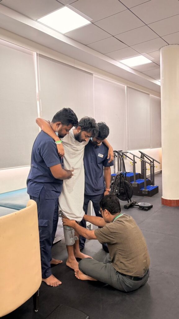
Paralysis, weakness, or numbness
loss of control over one’s bowels or bladder
Diagnosis
X-rays for preliminary evaluation
CT scans for the specifics of fractures
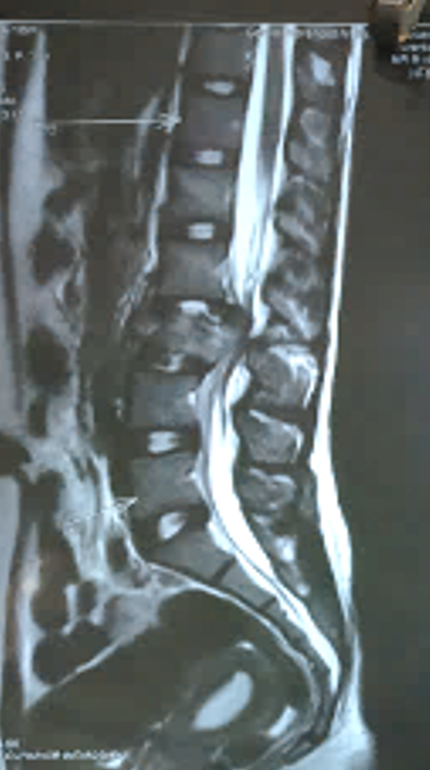
MRI to assess damage to the spinal cord and ligaments
Treatment
Conservative treatment using bracing and bed rest
Rehabilitation and pain management
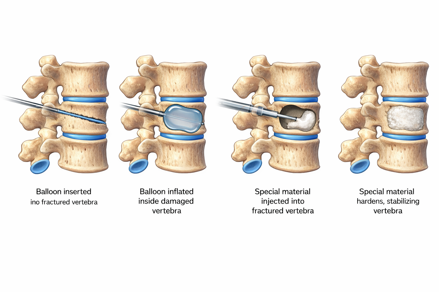
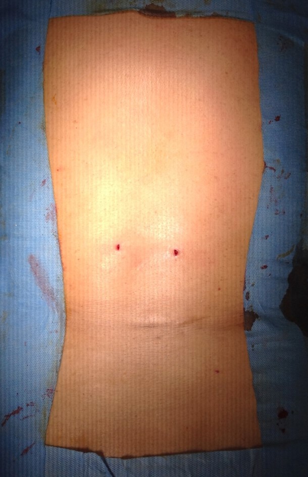
Minimally invasive treatments like kyphoplasty or vertebroplasty
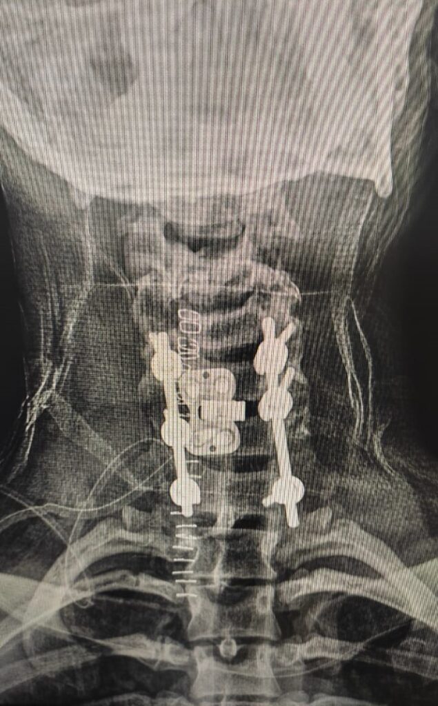
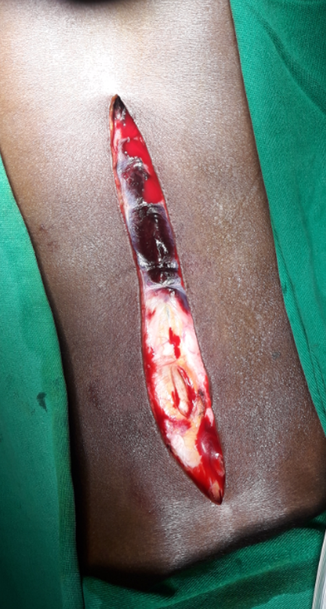
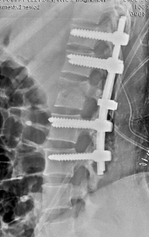
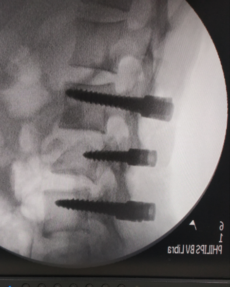
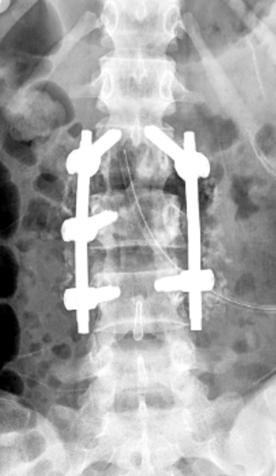
Surgical stabilization using cages, screws, or rods
1. Traumatic 2. Osteoporotic
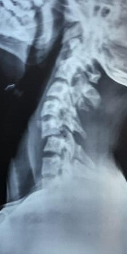
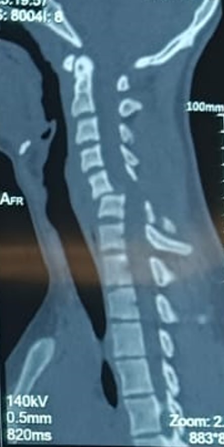
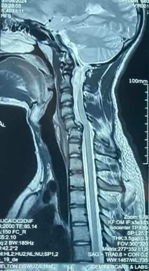
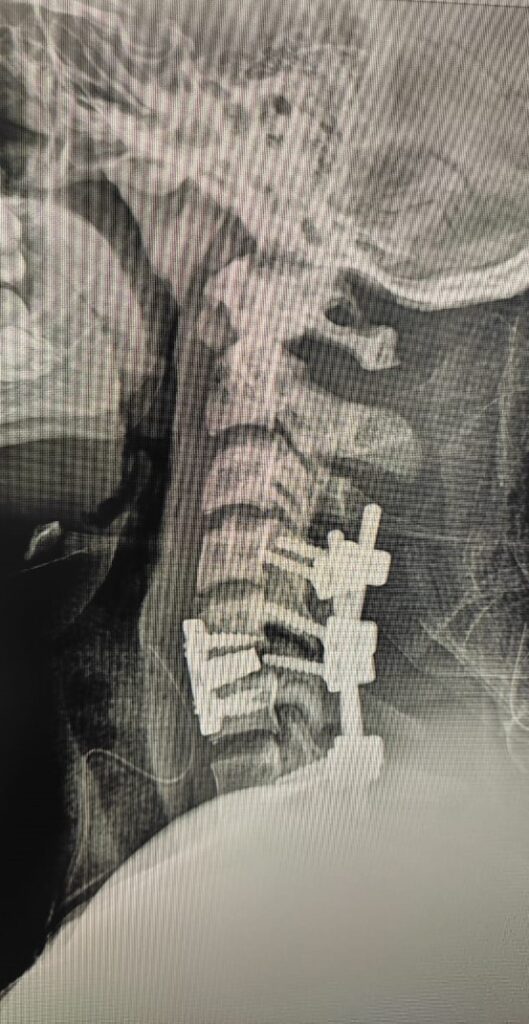
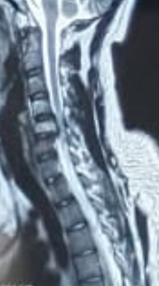
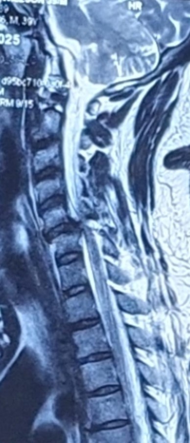
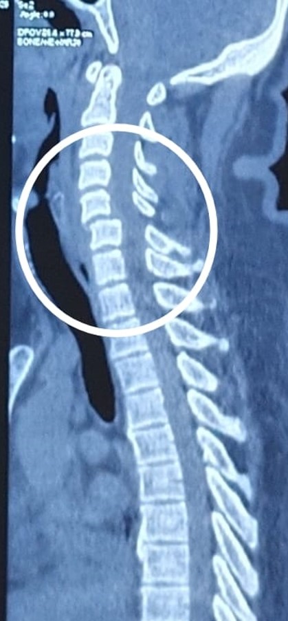
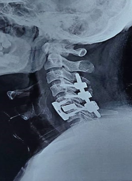
Traumatic Fracture dislocation C5-C6 with cord compression
Traumatic Burst Fracture C5 with ASIA A
Traumatic Fracture dislocation C5-C6 with cord compression and L1 burst fracture
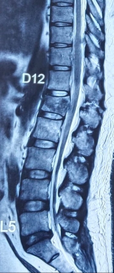
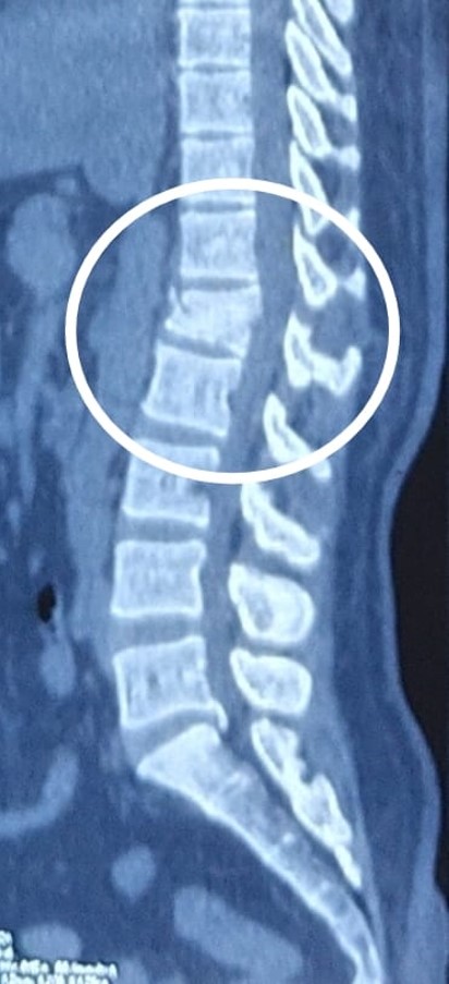
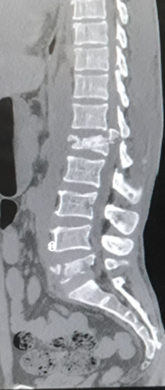
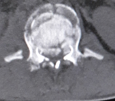
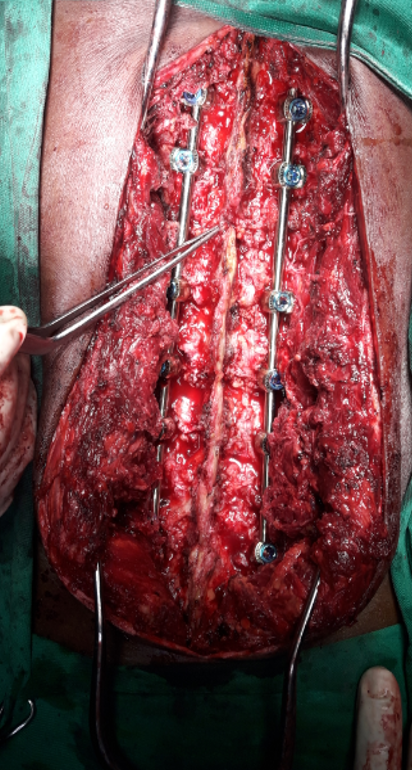
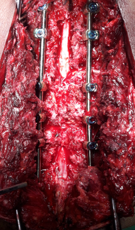
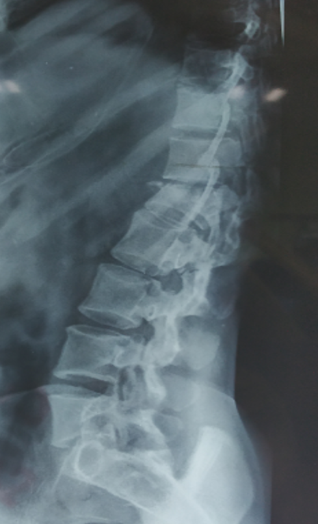
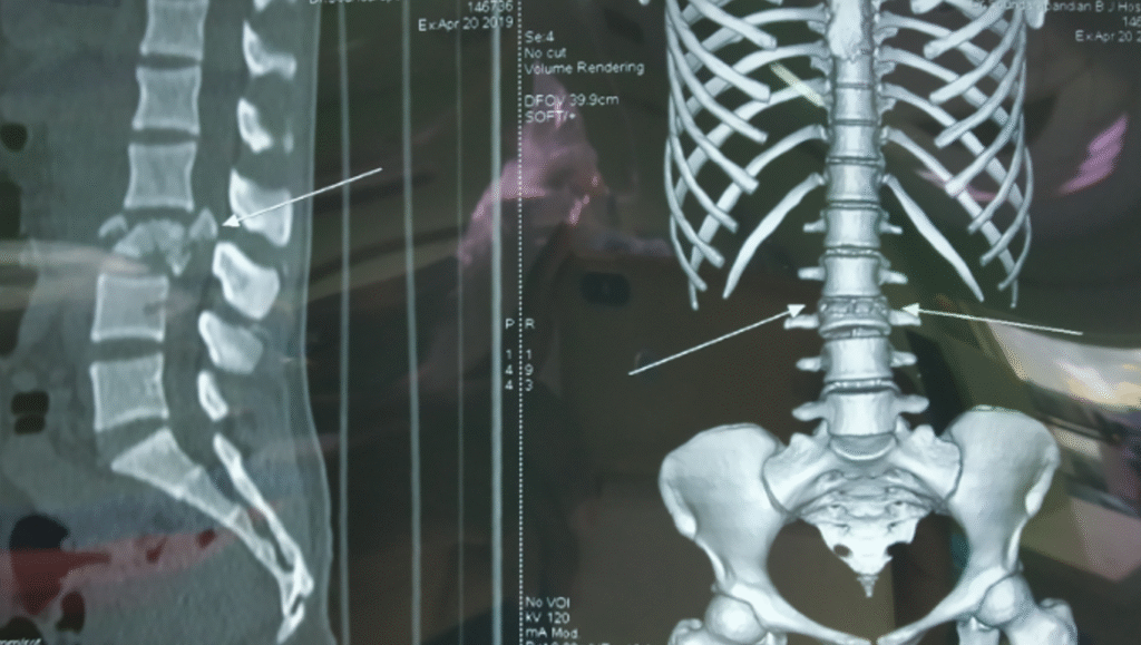
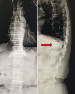
Burst Fracture L1 and L5 with retropulsion and neurological deficits
Burst Fracture L1 vertebra retropulsion and neurological deficits
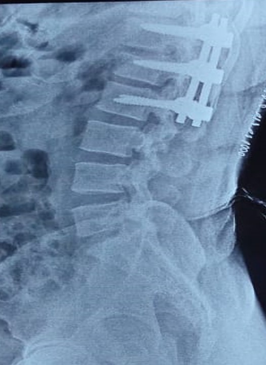
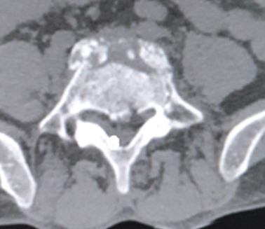
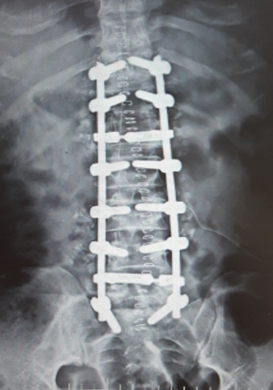
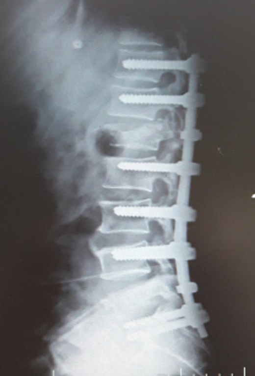
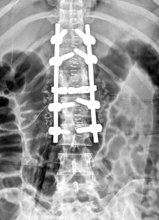
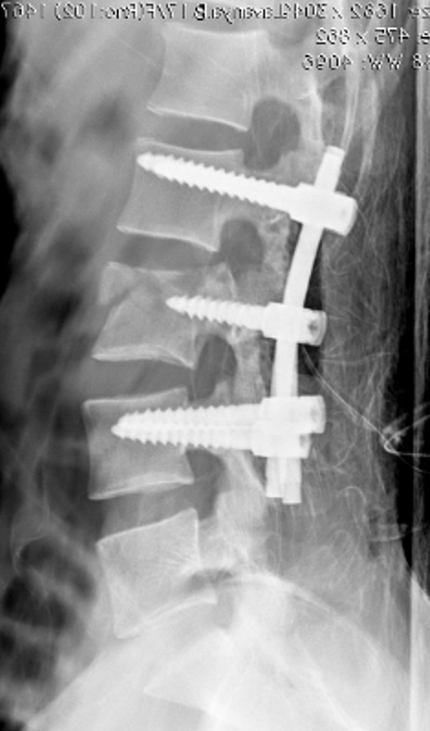
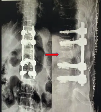
Post op x ray with local kyphosis correction and regain full vertebral height
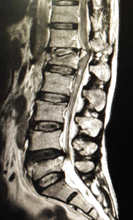
21 year girl Burst Fracture L3 with retropulsion and neurological deficits
Post op x ray with local kyphosis correction and regain full vertebral height
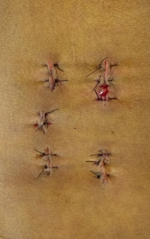
Minimal invasive spine surgery
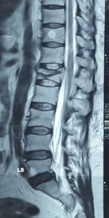
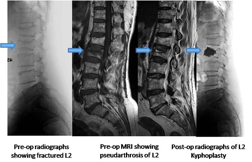
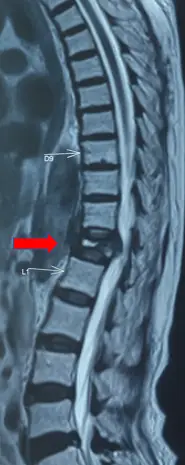
Osteoporotic Burst fracture L1
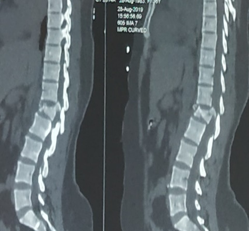
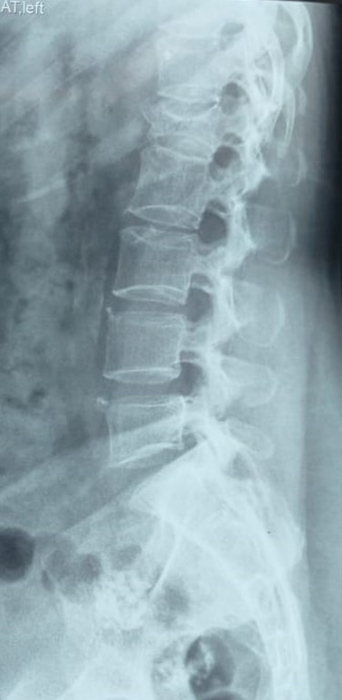
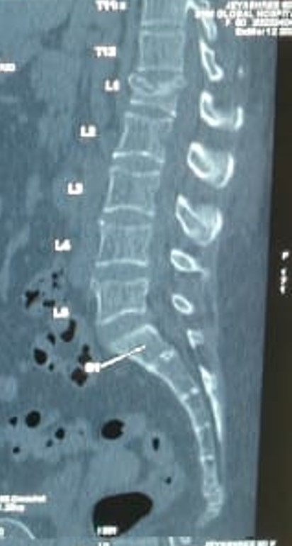
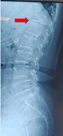
X ray imaging, CT scan and MRI of dorsolumbar spine showing osteoporotic vertebral compression fracture of more than 50 percent height loss at L1 vertebral body
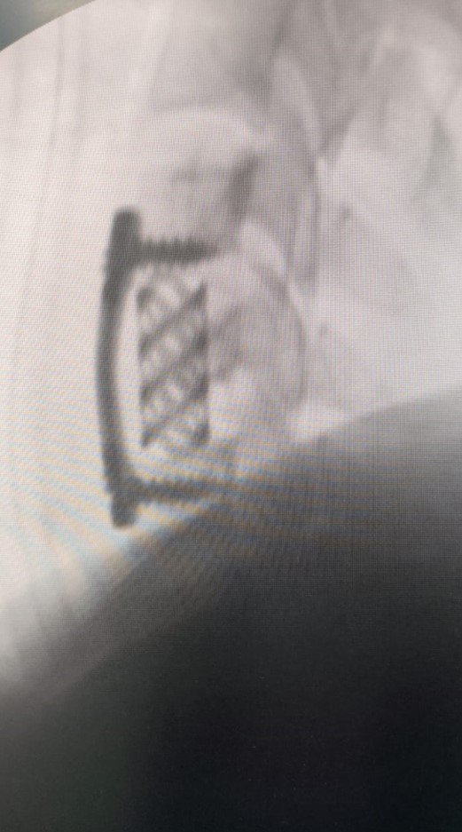
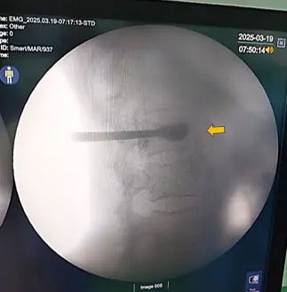
Intraoperative C-arm image showing Balloon dilatation by transpedicular jamshedi needle and restoration of height at L1 vertebral body
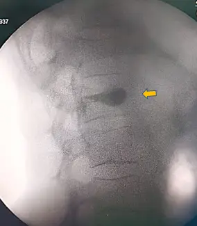
Final Intraoperative C-arm image showing full restoration of height with PMMA cement filling the void at the L1 vertebral body
Percutaneous Balloon Kyphoplasty
A 91 year Old active woman with osteoporotic compression underwent balloon kyphoplasty
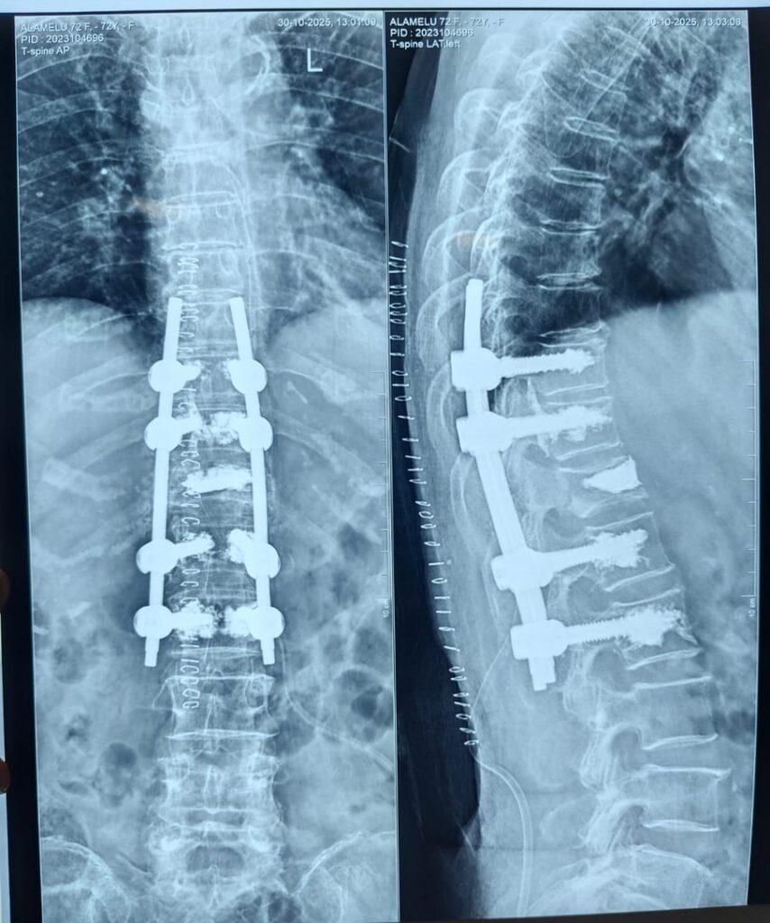
A 76 year Old woman with osteoporotic burst fracture and instability fixed with cement augmented screws
A 72 year Old woman with osteoporotic burst fracture / Parkinsons disease and bed ridden for 1 month